

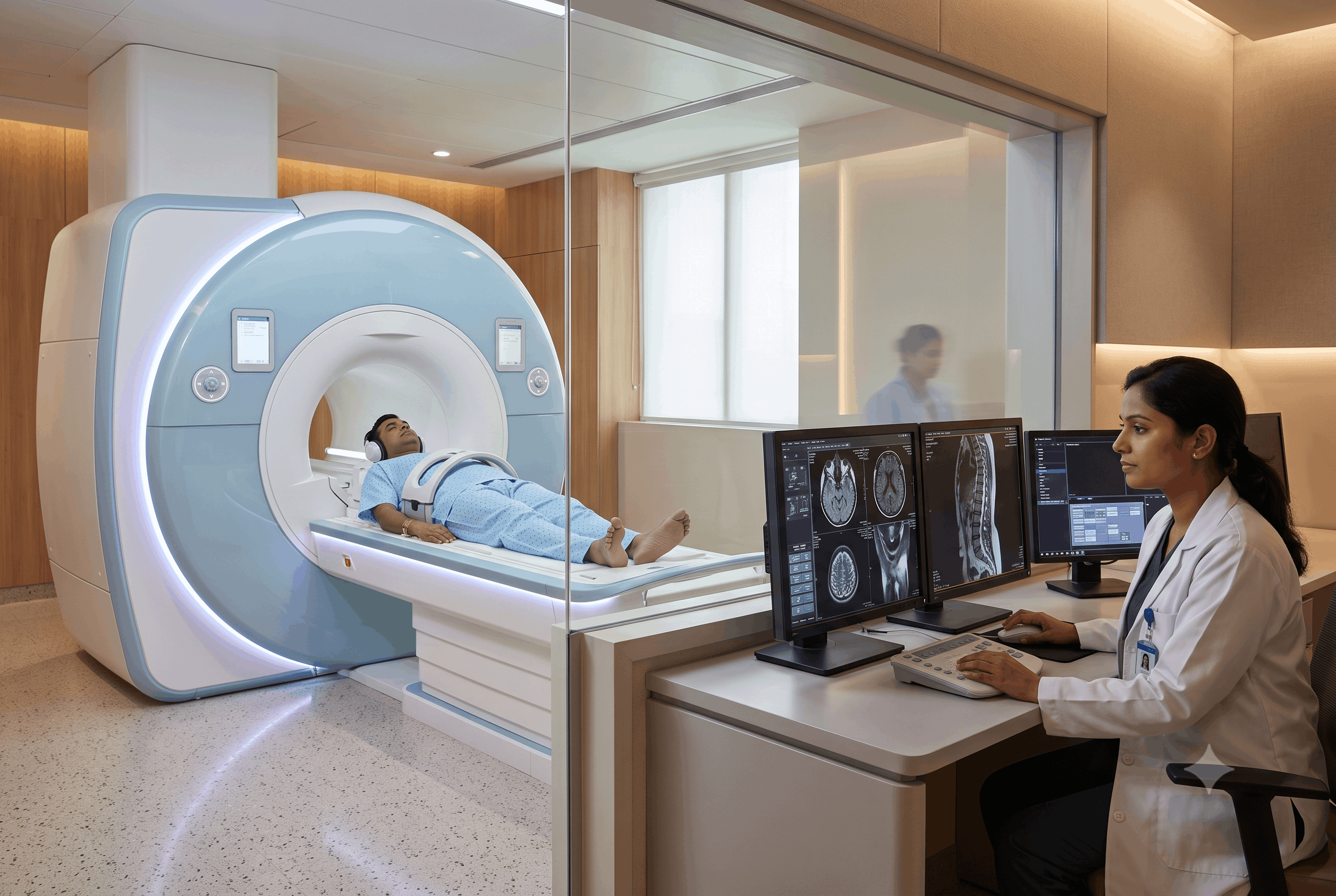
Early detection is key to improving the outcome of cancer treatment. If a person can be diagnosed with cancer before they have metastasised, they will usually have a more successful cancer treatment outcome and may require less aggressive treatment. Medical imaging technology has progressed rapidly in recent years, and new technologies are available to assist healthcare professionals in the earlier diagnosis of cancer. Whole body MRI technology, in particular, has been receiving a great deal of attention as a means of imaging many different organ systems in a single imaging session while not exposing a patient to any ionising radiation. While Whole Body MRI may help to pick up many types of cancer at an earlier stage than other modalities, Whole Body MRI is not a universal screening tool for the detection of cancers; the appropriateness of Whole Body MRI as a screening tool depends on an individual’s medical history, symptoms, and other individual risk factors related to their cancer development.
A Whole Body Magnetic Resonance Imaging (MRI) scan is a highly detailed imaging test that visualises most major organs and tissues in the entire body. Unlike traditional X-ray and CT imaging, an MRI uses very strong magnets and radio waves to acquire images instead of using ionising radiation.
During an MRI scan, images are taken of many different areas of the body, including the brain, neck, spine, chest, abdomen, pelvis, muscles, bones, and soft tissues. The scan typically takes anywhere from 45 to 90 minutes depending on the imaging protocol and whether contrast is needed.
MRI is a great tool to identify soft tissue abnormalities because MRI provides a great degree of soft tissue contrast.
Cancer is a condition that results from the abnormal growth and replication of cells resulting in mass formation and/or infiltration of surrounding tissue/masses. The reason MRI works well to detect cancer is that no two tissues will produce the same signals when exposed to a magnetic field, making it very easy for radiologists to identify “suspicious abnormalities”.
Possible abnormal findings on a Whole Body MRI could include:
MRI is not used to directly diagnose cancer but only to find “suspicious” findings that require additional testing (imaging or laboratory).
Whole Body MRI has been proven effective at finding many types of cancer, especially cancers that arise from soft tissue.
Examples:
MRI can help to find abnormalities in both the bone marrow and surrounding soft tissue. In addition, it will help to find primary bone cancer and also cancers that have spread to bone from other areas of the body.
MRI can show very detailed imaging of liver tissue and is most often used to detect lumps or abnormal areas suspected to be liver cancer, as well as to evaluate abnormal liver-type lesions and monitor those patients that have chronic liver disease.
Kidney tumours are often asymptomatic in the early stages of development, so it is difficult to determine that a person has a kidney tumour until it is advanced. MRI will help to find kidney masses but can also help to differentiate between benign and malignant tumours.
Pancreatic cancer has one of the lowest incidences of producing an early diagnosis. MRI provides excellent evaluation of the pancreas and surrounding tissues when abnormalities are suspected.
Whole body MRI protocols may discover a primary brain tumour with sufficient accuracy to warrant further follow-up; however, in a suspected case of brain tumour where there are neurological findings, a dedicated Brain MRI will provide the greatest accuracy.
Because of the ability to identify bone marrow involvement in a patient with MM throughout the skeletal system, it is becoming the preferred imaging method for patients with MM when compared to conventional radiography.
MRI is recognized as the best imaging modality to visualize tumours that start in muscle, fat, nerve, and connective tissue.
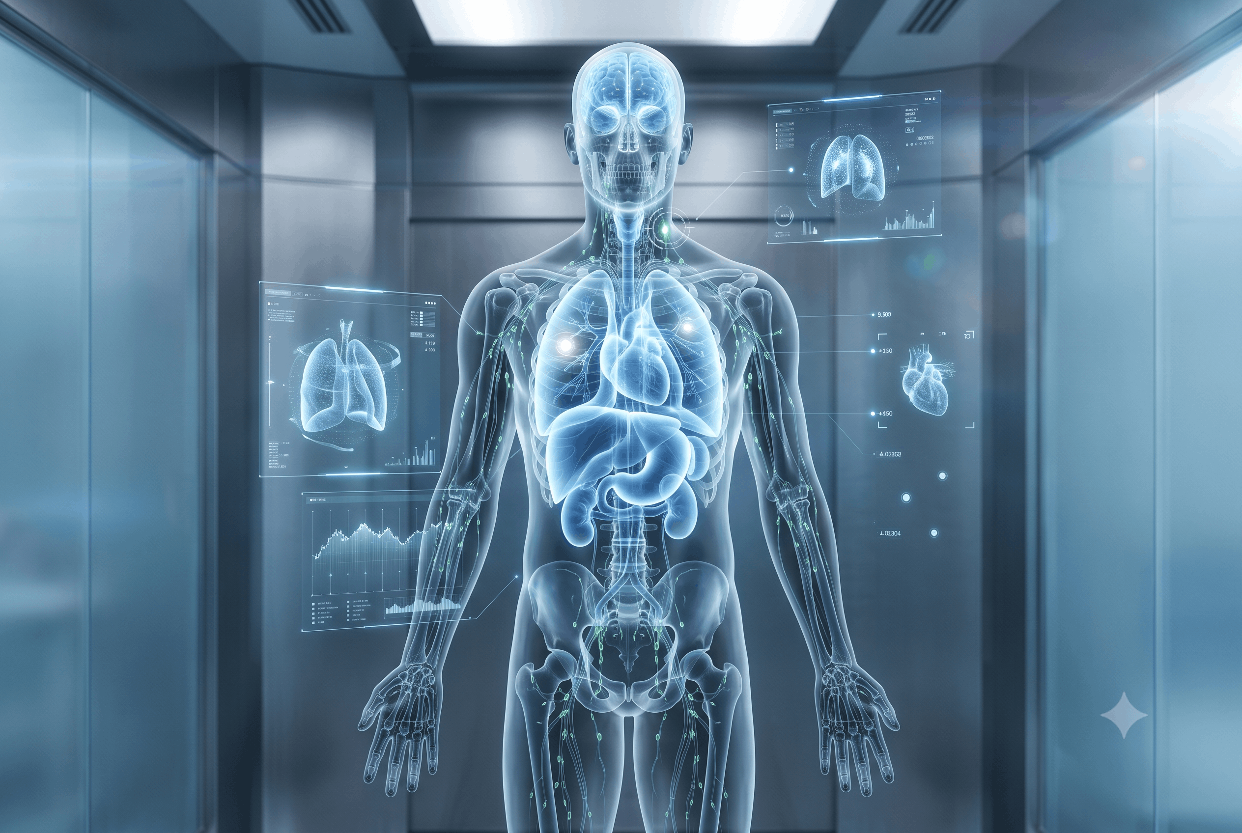
Whole Body MRI is a very good diagnostic tool, but it’s not 100% accurate, and there are many types of cancer that it cannot identify.
Examples of limitations include:
For these reasons, Whole Body MRI should not replace standard cancer screening tests such as mammography, colonoscopy, cervical cancer screening or low dose CT screening on individuals with multiple risk factors for lung cancer.
Whole body MRI is not justified as a routine screening exam for the general population; however, it may be justified in select patients.
Possible candidates include:
Patients should always discuss with their physician whether or not a whole body MRI is appropriate.
Unlike CT imaging, a Whole Body MRI does not expose patients to ionizing radiation, making it an option for those needing repeated imaging.
MRI provides much better visualization of organs, muscles, nerves, blood vessels, and connective tissue, and is therefore particularly effective for detecting abnormalities in the soft tissues.
Rather than simply examining a single organ, Whole Body MRI allows for multiple body regions to be evaluated during one scan.
In addition to identifying cancer, Whole Body MRI can also help detect other disease types, such as inflammatory disease, vascular abnormalities, spinal disorders, and musculoskeletal issues.
Whole Body MRI has its limitations, just like other diagnostic tests.
There could be incidental findings on the scan that would not have anything to do with the patient’s symptoms. Although most of these findings will not result in finding out anything significant, they may result in further testing, follow-Up imaging or a biopsy to determine if there is any clinical value to the incidental finding.
Some patients will not be able to tolerate the enclosed scanner due to claustrophobia or will find it uncomfortable due to the length of time they are in the scanner, however Modern MRI technology includes features that are designed to help reduce the impact of claustrophobia and discomfort during scanning. Medical teams are also able to help reduce anxiety for patients during the scan.
Neither type of imaging is better than the other in all settings. While CT imaging is the preferred type of imaging for emergency trauma patients; when looking at the lungs for a lung issue; when looking for fractures; and for the rapid assessment of acute medical conditions, MRI imaging is superior when looking at soft tissues, the brain, spinal cord, joints, muscles, and most of the internal organs.
Medical professionals will use the patient’s presenting symptoms and medical history to determine which type of imaging test is most appropriate to help answer the specific clinical question being asked.
The issue of whether healthy individuals benefit from whole body MRIs continues to be a controversial topic among medical experts worldwide.
A small number of individuals may choose to have a Whole Body MRI as an initial preventive health screening test because they possess multiple risk factors for developing cancer and/or have a positive family history of hereditary cancer predisposal syndromes. However, due to the lack of sufficient scientific research on the overall benefit, cost-effectiveness, and impact of Whole Body MRIs on long-term patient health outcomes, the majority of professional medical organizations do not recommend performing Whole Body MRIs on a routine basis as part of a health screening for the general population.
Instead, it is recommended that patients obtain routine health evaluations through their primary care providers, along with participating in age-appropriate and risk-based screenings that are currently recognized as standard-of-care in clinical practice.
The whole body MRI is considered one of the most advanced technologies currently used in the detection of diseases, including some types of cancer. Because it yields high-quality and detailed images of several different organs without exposing patients to radiation, the whole-body MRI can be very helpful in specific groups of patients with an elevated risk of developing cancer or having symptoms that remain unexplained. While helpful in some patients, the whole body MRI cannot replace traditional screening methods and it will not detect all types of cancer.
The best way to detect cancer early is to have a combination of symptom identification, healthy lifestyle choices, regular medical exams, recommended screening tests, and then when indicated by a medical professional, appropriate imaging. Aarna Diagnostic Centre in Dehradun can be a resource for patients who may need advanced MRI technology as part of a comprehensive evaluation.
Convenient Tests right at your Doorstep!


86, Kargi – Patel Nagar Bypass, near Palm City, Adarsh Vihar, Brahmanwala, Dehradun, Uttarakhand 248001
1, Maharani Bagh Lane, Ballupur Chowk, Ashirwad Enclave, Ballupur, Dehradun, Uttarakhand 248001
1, Chakrata Rd, near Rohan Motors, Selakui, Central Hope Town, Uttarakhand 248011
F-7, Haridwar Road Nehru Colony Dehradun, Uttarakhand 248001 India
Leave a Comment