

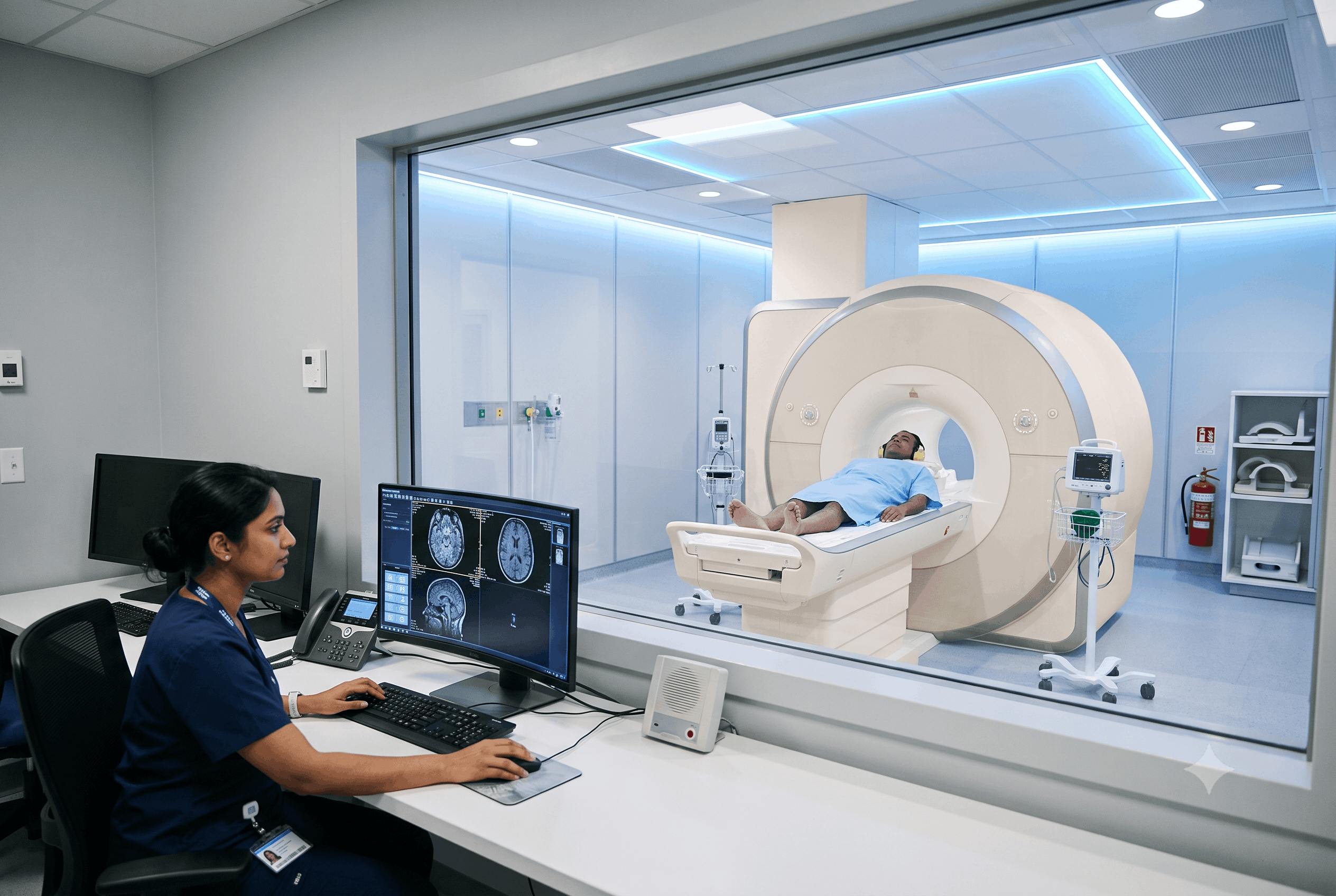
Magnetic Resonance Imaging (MRI) is one of the most advanced diagnostic imaging techniques used in modern healthcare. It uses a powerful magnetic field and radio waves to produce highly detailed images of the body’s internal structures without exposing patients to ionising radiation. Because of its exceptional ability to visualise soft tissues, MRI plays a vital role in identifying a wide range of medical conditions affecting the brain, spine, joints, muscles, blood vessels, and internal organs. Understanding the common conditions diagnosed through MRI helps patients appreciate why doctors often recommend this scan when precise imaging is needed. In many situations, MRI provides greater detail than other imaging methods, supporting accurate diagnosis, effective treatment planning, and improved patient care.
Neurological disorders are one of the most frequent applications of MRI. The high quality and clarity of brain tissue images created by MRI make it a valuable tool for detecting abnormalities that may not be apparent in other imaging techniques. Brain tumors are one of the types of abnormalities often detected through MRI. The MRI helps determine the tumor’s size, location, and characteristics—all necessary for proper treatment planning—because it can detect any changes to the surrounding brain tissue. Patients seeking positive identification of a neurologically-based cause for persistent headaches or other neurological symptoms like seizures, unexplainable dizziness, weakness, memory loss, or symptoms affecting their ability to see, often undergo MRIs.
Stroke detection is another important application of MRI. MRI can identify whether there has been an area of brain tissue that was damaged or is in the process of being damaged from a lack of adequate blood flow. An MRI examination can also identify very early changes in brain tissue before significant damage occurs. MRI can help diagnose multiple sclerosis, a condition that damages the protective sheath around nerve fibres, as well as infections; inflammation; congenital abnormalities of the brain; and certain types of dementia.
The MRI is one of the best ways to see the spine in detail, due to its ability to demonstrate the spinal cord, discs, nerves, muscles, and other soft tissues surrounding the spine with great clarity.
An example of a very common spinal condition diagnosed by MRI is the herniated or “slipped” disc. A herniated disc occurs when the soft, inner portion of a disc between two vertebrae pushes through the tough outer layer of the disc and may press against adjacent nerves, causing pain, numbness, or weakness in specific areas of the body. An MRI helps determine the exact location and degree of this problem.
Spinal conditions that are regularly diagnosed with MRI include spinal stenosis, degenerative disc disease, spinal cord compression, infections, tumours, fractures of soft tissues and inflammatory conditions.
MRIs are particularly helpful in the diagnosis of patients who have chronic neck pain, low back pain, or sensations of tingling or weakness to their arms or legs.
The MRI is an important tool for the orthopaedic medical physician; the MRI is extremely good at imaging the soft tissue associated with the joints of the human body.
The MRI is frequently used to diagnose meniscus (cartilage) tears, ligament injuries (anterior cruciate ligament, or ACL, tears), cartilage injury, tendon injuries, and bone marrow abnormalities in the knee.
While there are other expected uses of an MRI in the knee, there are also expected uses for the MRI in diagnosing similar joint injuries in the shoulder, hip, ankle, wrist, and elbow.
Many common sports injuries affect muscle, tendon and ligament structures; however, standard X-ray imaging does not provide sufficient information about these injuries. The use of magnetic resonance imaging (MRI) provides the required state-of-the-art imaging capabilities to help doctors diagnose and determine the appropriate treatment course (conservative therapy, physiotherapy or surgical intervention) for an injured patient.
MRI can help to find abnormalities in both the bone marrow and surrounding soft tissue. In addition, it will help to find primary bone cancer and also cancers that have spread to bone from other areas of the body.
MRI can show very detailed imaging of liver tissue and is most often used to detect lumps or abnormal areas suspected to be liver cancer, as well as to evaluate abnormal liver-type lesions and monitor those patients that have chronic liver disease.
Kidney tumours are often asymptomatic in the early stages of development, so it is difficult to determine that a person has a kidney tumour until it is advanced. MRI will help to find kidney masses but can also help to differentiate between benign and malignant tumours.
Pancreatic cancer has one of the lowest incidences of producing an early diagnosis. MRI provides excellent evaluation of the pancreas and surrounding tissues when abnormalities are suspected.
Whole body MRI protocols may discover a primary brain tumour with sufficient accuracy to warrant further follow-up; however, in a suspected case of brain tumour where there are neurological findings, a dedicated Brain MRI will provide the greatest accuracy.
Because of the ability to identify bone marrow involvement in a patient with MM throughout the skeletal system, it is becoming the preferred imaging method for patients with MM when compared to conventional radiography.
MRI is recognized as the best imaging modality to visualize tumours that start in muscle, fat, nerve, and connective tissue.
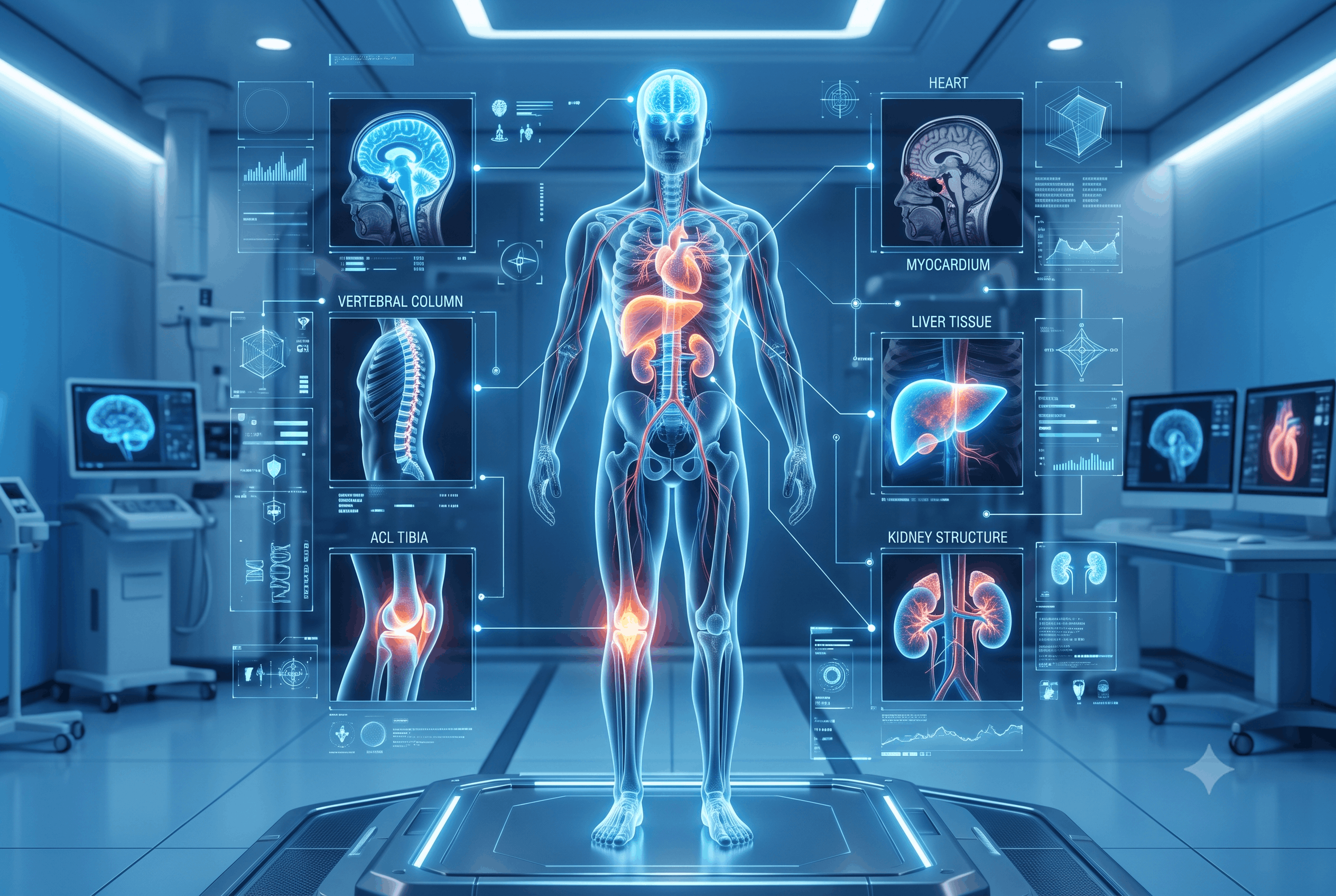
Although X-ray imaging will continue to provide the first-line imaging method for the majority of fractures, MRI can also provide additional diagnostic information regarding certain types of bone abnormalities that may not be identified through conventional imaging. Bone fractures (stress fractures), infectious processes involving the bone, bone marrow pathologies, and early signs of osteonecrosis are just some examples of bone medications that can be diagnosed using MRI. MRI is also very useful in assessing muscle, tendon and soft tissue pathologies :including muscle ruptures, tendonitis/tendon inflammation, soft tissue septic and chronic muscle pain.
If a patient continues to have pain following an injury and appropriate testing has not identified the cause of this pain, an MRI may be ordered.
MRI is also now becoming an increasingly common modality for assessing cardiovascular diseases. MRI helps doctors evaluate heart muscle (myocardium) function, assess heart chamber dimensions, evaluate congenital heart disease, evaluate heart inflammation, cardiomyopathy & select valve disorders.
MRI provides a complete vascular assessment by creating detailed images of all major blood vessels to assist in the identification of aneurysms, vascular malformations, and arterial stenosis.
There are clinical situations when it may be appropriate to use MRI in conjunction with other cardiac imaging modalities depending on the pathology being evaluated.
MRI is often used to assess abdominal organs specifically when the information obtained from either ultrasound or CT requires clarification.
Liver imaging can be used to investigate liver tumors, fatty liver disease, cysts, cirrhosis, and other liver abnormalities. Other abdominal organs that may be assessed in addition to the liver include pancreas, kidney, gallbladder, spleen, adrenal, and bile ducts.
MRI can also be recommended for individuals with continuing abdominal pain, unintentional weight loss, abnormal liver function testing, or those with an abnormality that is suspected to be a tumor.
MRI also helps with the evaluation of numerous kidney disorders, including kidney tumors, cysts, congenital disorders, and urinary tract obstructions.
Additionally, individuals with contraindications for contrast-enhanced CT scans or who require more precise soft tissue assessments may be referred for an MRI of their kidneys and urinary tracts.
MRI allows for detailed evaluation of the surrounding organs and structures to assist physicians in determining the extent of disease.
The use of MRI in gynecology is rapidly becoming an invaluable diagnostic tool due to the high-quality imaging it provides of the pelvic organs.
MRI is often the preferred imaging modality for the evaluation of uterine and ovarian fibroids (cysts), endometriosis, congenital uterine anomalies, and select pelvic tumours. In addition, MRI can help assess the presence of any unclear ultrasound findings in assessing complex pregnancy-related conditions.
Breast MRI is a further use of MRI in medicine; it is a diagnostic tool used in carefully selected patients (primarily those at a high risk of breast cancer or those requiring additional evaluation after an abnormal mammogram and/or ultrasound).
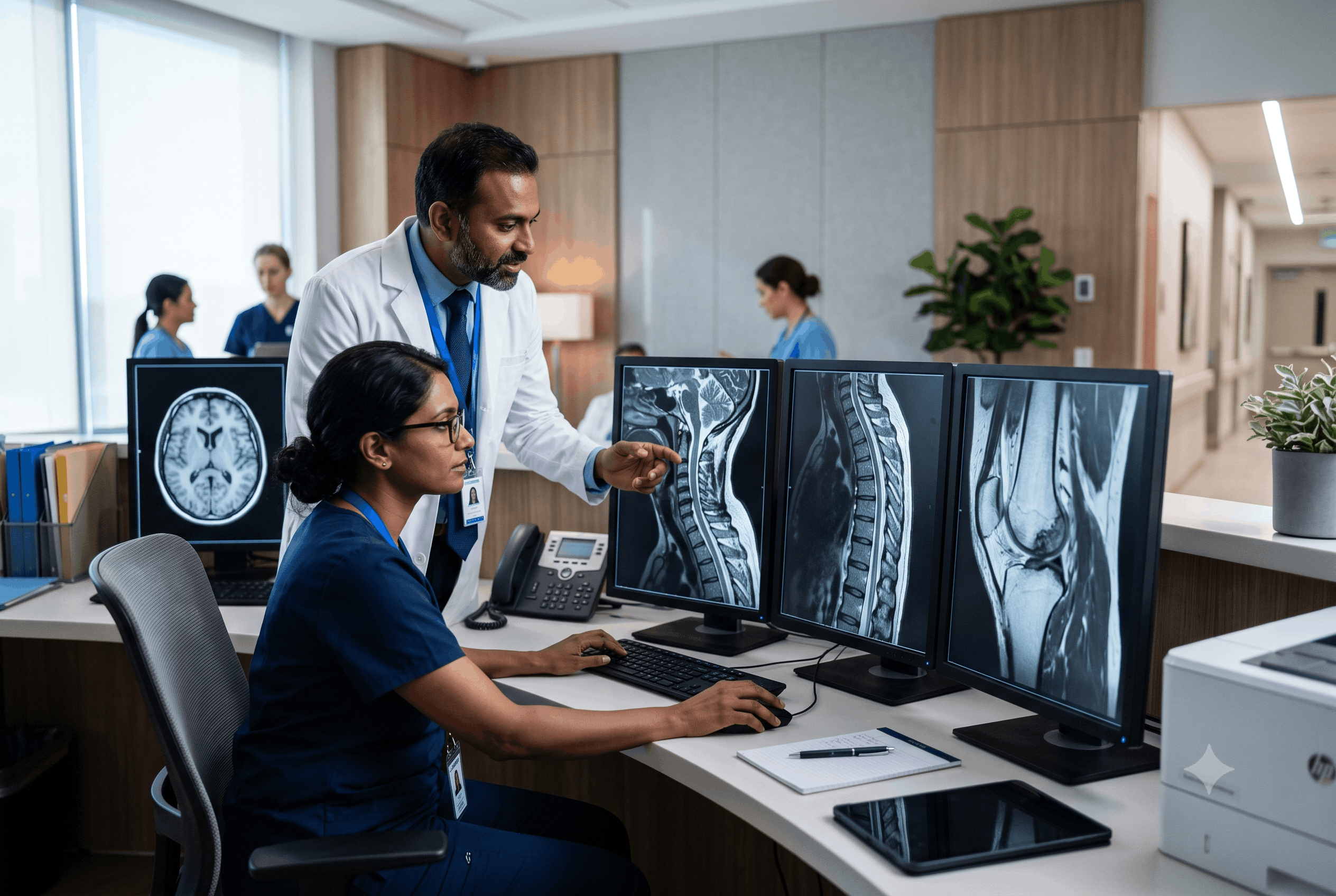
MRI is used extensively in oncology, particularly because it can provide highly detailed images of soft tissues.
Although MRI alone cannot definitively diagnose if a mass is malignant or benign, it is critical to help identify masses that are suspicious for malignancy, their relative sizes, whether or not they are impinging on adjacent structures, and how well they are responding to treatment.
MRI is most commonly performed on brain, spine, liver, prostate, uterus, breast, pancreas, kidney, and musculoskeletal tumours. It also has a key role in evaluating follow-up images after surgical resection, chemotherapy, or radiation therapy.
In today’s society people suffer from a variety of disorders including autoimmune and inflammatory disorders. There are multiple types of inflammatory diseases such as joint inflammation, soft tissue inflammation, muscle inflammation and nerve tissue inflammation. MRI allows for the detection of these disorders in their earlier stages so that early intervention/treatment can be provided.
Examples of autoimmune diseases where MRI can be helpful include rheumatoid arthritis, multiple sclerosis, inflammatory bowel disease, inflammatory muscle disease, and autoimmune diseases affecting the brain or spinal cord.
By diagnosing these autoimmune and inflammatory diseases early there is an opportunity for medical professionals to initiate treatment prior to irreversible damage occurring.
There are several features of MRI that make it a more suitable imaging modality over other imaging modalities. The most significant advantage is the ability of MRI to provide excellent contrast between different types of soft tissue. As a result, doctors are able to accurately delineate between multiple types of soft tissue.
Another advantage of MRI is that it does not use ionising radiation. Therefore, it is a safe alternative to many patients who need to be imaged multiple times over their lifetime.
MRI has the additional advantage of imaging in numerous planes of view without repositioning a patient. Consequently, radiologists are able to look at organs and tissues from multiple angles which increases the accuracy of the radiologists’ diagnoses.
MRI will not always be the first imaging modality chosen to assess a given patient’s condition. Factors like the patient’s presenting symptoms, medical history, urgency of the condition, presence of conductive devices in the patient’s body, and area of the body being examined all affect which imaging modality will be chosen.
MRI has transformed the way that many illnesses are diagnosed by providing images of detailed, high-quality pictures of body tissue such as brain, spine, and muscles, to name a few. As a result, doctors utilize MRI technology to ascertain the presence and/or severity of conditions such as brain injury, spine injury, sports injury, coronary artery disease, abdominal disease, or cancer, among other things, with a high degree of fidelity. MRI technology can usually be used as an adjunct to determine the diagnosis, severity of a condition, and/or to plan for the future course of treatment (i.e., to provide an accurate picture).
Patients should understand what types of conditions are detectable via MRI imaging so that they can become educated about decisions related to their health care. When a qualified health care professional determines that an MRI is warranted, scheduling an MRI appointment and completing the MRI scan may lead to an early diagnosis of a medical condition, providing a more effective course of treatment, and improving overall health outcomes. For example, the availability of Aarna Diagnostic Centre’s MRI facility in Dehradun, along with other MRI facilities, provides Dehradun residents access to state-of-the-art diagnostic imaging, if warranted.
Convenient Tests right at your Doorstep!


86, Kargi – Patel Nagar Bypass, near Palm City, Adarsh Vihar, Brahmanwala, Dehradun, Uttarakhand 248001
1, Maharani Bagh Lane, Ballupur Chowk, Ashirwad Enclave, Ballupur, Dehradun, Uttarakhand 248001
1, Chakrata Rd, near Rohan Motors, Selakui, Central Hope Town, Uttarakhand 248011
F-7, Haridwar Road Nehru Colony Dehradun, Uttarakhand 248001 India
Leave a Comment