

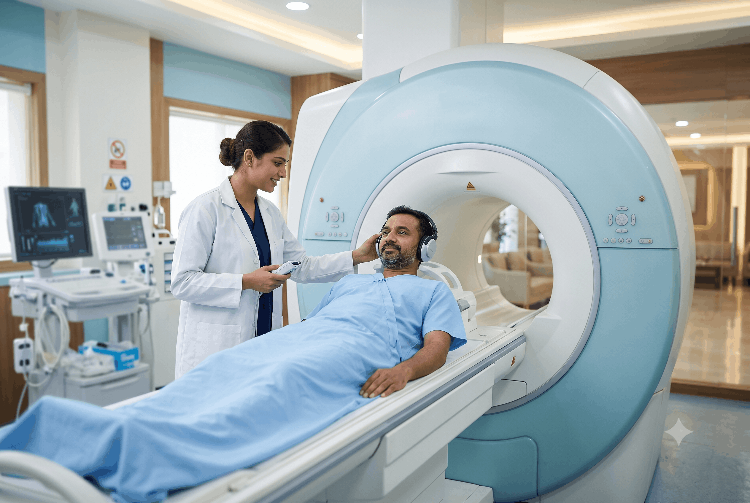
As a result of advances in medical imaging, there has been a dramatic shift in how physicians diagnose diseases and injuries. Physicians no longer have to rely solely on physical examinations or invasive procedures; they can now view the patient’s internal anatomy with extreme precision through a variety of imaging techniques. MRI (magnetic resonance imaging) is one of the more advanced imaging modalities, producing highly detailed images without exposing patients to ionizing radiation.
To reduce anxiety and assist in providing timely diagnoses when a healthcare professional recommends an MRI, potential patients should understand how MRIs work, the reason for the MRI, and what they can anticipate when completing an MRI.
MRI (Magnetic Resonance Imaging) is a method of diagnostic imaging that provides detailed “cross-sectional” images of the internal structures of the body utilizing a large, powerful magnetic field, radiofrequency (RF) signal and computer-based technology to generate the images. MRI uses no form of ionizing radiation; it relies on the intrinsic nature of hydrogen atoms that occur in the water molecules in the body.
Due to the high-water content of the human body, MRI is an excellent way to see soft tissue structures, such as the brain, spinal cord, muscles, ligaments, cartilage, blood vessels, and internal organs. By showing an incredible amount of detail, MRI can often provide significant help to physicians in diagnosing certain conditions that may not be as visible through other imaging modalities.
The underlying science of MRI may seem complicated, but the underlying principle is simple.
An MRI scanner contains a very powerful magnet, used to cause hydrogen atoms in the body to align with the magnetic field of the MRI. Once these hydrogen atoms are aligned, the MRI scanner then sends very precisely controlled radiofrequency energy pulses to disrupt the alignment of these hydrogen atoms. As the hydrogen atoms return to their pre-disruption position, they create very small signals.
These signals are detected by specialized sensors in the MRI scanner. Millions of these signals are processed using sophisticated computer software applications, generating very detailed images of different body tissues.
Different tissues within the human body contain different quantities of water, producing different signalling patterns. Based upon this, radiologists can easily identify healthy tissue from non-healthy tissue with incredible accuracy.
The use of an MRI is extensive and can be utilized to diagnose a multitude of medical issues among many specialties.
An MRI is extremely valuable for evaluating conditions that exist within the neurological realm. Some examples include:
An MRI may be the preferred imaging test for patients with prolonged headaches, seizures, loss of coordination, weakness that cannot be explained, or changes to vision.
An MRI will produce images of the spinal cord, vertebral column (backbone), and soft tissue surrounding them. An MRI is often performed to evaluate:
An MRI is frequently used by physicians specializing in sports medicine and orthopedics, as it clearly shows soft tissue injuries to:
An MRI can identify soft tissue injuries when other imaging techniques may not.
An MRI can help evaluate benign or malignant tumors, cysts, and chronic inflammatory conditions of the following organs:
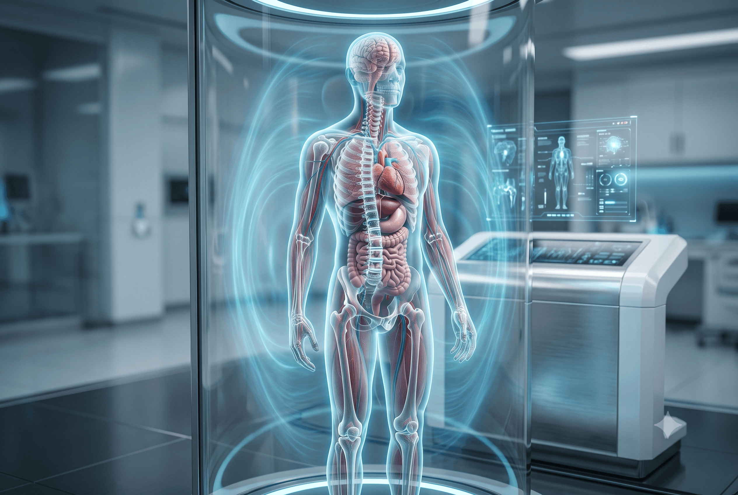
Specialised cardiac MRI studies help evaluate:
The majority of physicians will use an MRI to see clearly the anatomy of soft tissues. If previous radiologic studies didn’t provide enough information, then MRI is usually utilized.
Examples of this would be:
Symptoms that could lead a physician to consider an MRI include:
The decision to proceed with an MRI is based upon the patient’s presenting clinical symptoms, the physician’s or qualified health care professional’s assessment of the patient’s medical history or clinical assessment.
Before having an MRI, a lot of people feel anxious; the main reason is that they do not know what will be done to them. The actual procedure is non-invasive and generally enjoyable.
Before the MRI starts, patients must remove all metallic items, including rings, watches, and hearing aids. The reason for this is that the MRI machine contains a strong magnet.
The patient is positioned on a movable table that moves slowly into the MRI machine. The MRI machine makes loud noises while taking the actual pictures; staying as still as possible is essential in order to obtain clearer images.
Generally, patients will be given earplugs or headphones to listen to while trying to relax.
Depending on the body part being examined, an MRI scan typically takes between 20 and 60 minutes.
Some MRI exams can have the quality of the picture improved when the use of a contrast agent is made; the contrast substance, which is usually made up of gadolinium, is injected either before or at some point during the imaging arousal, through the vein.
Contrast-enhanced MRI allows doctors and other health care professionals to better visualise:
Not every MRI requires the use of contrast material. The use of contrast will only be determined by the clinical question to be investigated.
MRI is considered one of the safest imaging modalities because it does not involve exposure to ionising radiation.
Nonetheless, while being very safe for the majority of people, there are specific cautionary measures. A patient needs to immediately let their health care provider be informed, prior to their MRI, if they have:
The majority of advances in implanted devices are that they are now considered to be compatible with MRI; however, in order to have genuine compatibility with MRI, they must be assessed prior to MRI.
It is also very important for any woman that is pregnant, that she informs her Doctor that she is pregnant prior to undergoing a MRI. Even though evidence indicates that MRI is a generally safe modality during pregnancy when needed statistically there may still be both benefits and risks incurred by the individual receiving an MRI.
Both MRI and CT scans are important diagnostic tools. However, they have different purposes.
CT Scans are X-ray based and are great for evaluating fractures, disorders of the chest, and identifying signs of internal bleeding or trauma resulting from an emergency situation due to injury, fall, etc. They also have the advantage of being available quickly and widely.
MRI scans provide greater detail for nerves, soft tissues, muscles, ligaments, and the brain. When more detailed examinations of soft tissue are needed, MRI is often the imaging technique of choice.
Neither type of scan is “better” than the other. Each has its strengths, and physical examination findings and/or clinical presentation will determine the most appropriate imaging to be done by the physicians to evaluate the patient.
Most importantly, the timely performance of imaging can sometimes prevent delays in diagnosis and/or treatment, so any new onset of symptoms that are persistent (e.g., headache, generalized weakness, back pain, recurrent seizures, persistent joint pain) should be evaluated by a healthcare provider. When an MRI scan is ordered, it should be done in a timely manner, which will allow identification of the cause of the symptoms.
The advances in MRI technology have made it possible to detect many conditions much sooner than previously, often well before significant complications occur. Early diagnosis usually allows for better planning and more effective treatment with better long-term outcomes.
To summarize, MRI has become one of the key tools available for diagnostic purposes in today’s modern medical world because of its non-invasive nature, as it allows physicians to obtain unprecedented levels of detail with no exposure to radiation. Moreover, the use of MRIs for evaluating neurological conditions, spinal issues, joint injuries, and the assessment of internal organs and possible disease processes has supported the physician’s ability to establish accurate medical diagnoses.
Having a basic understanding of how MRI works can assist individuals undergoing this procedure by providing information that will make them more knowledgeable and confident about their procedure. In addition, MRIs are an important part of identifying the underlying causes of ongoing symptoms and tracking ongoing condition(s). Therefore, MRI continues to provide an essential resource for the early diagnosis, timely initiation of treatment, and delivery of the best patient care. Through the presence of modern MRI facilities in Dehradun such as Aarna Diagnostic Centre and others, individuals whose physicians have recommended advanced imaging will have access to a valuable resource.
Convenient Tests right at your Doorstep!


86, Kargi – Patel Nagar Bypass, near Palm City, Adarsh Vihar, Brahmanwala, Dehradun, Uttarakhand 248001
1, Maharani Bagh Lane, Ballupur Chowk, Ashirwad Enclave, Ballupur, Dehradun, Uttarakhand 248001
1, Chakrata Rd, near Rohan Motors, Selakui, Central Hope Town, Uttarakhand 248011
F-7, Haridwar Road Nehru Colony Dehradun, Uttarakhand 248001 India
Leave a Comment